Contrary to some reports, you don't have two brains
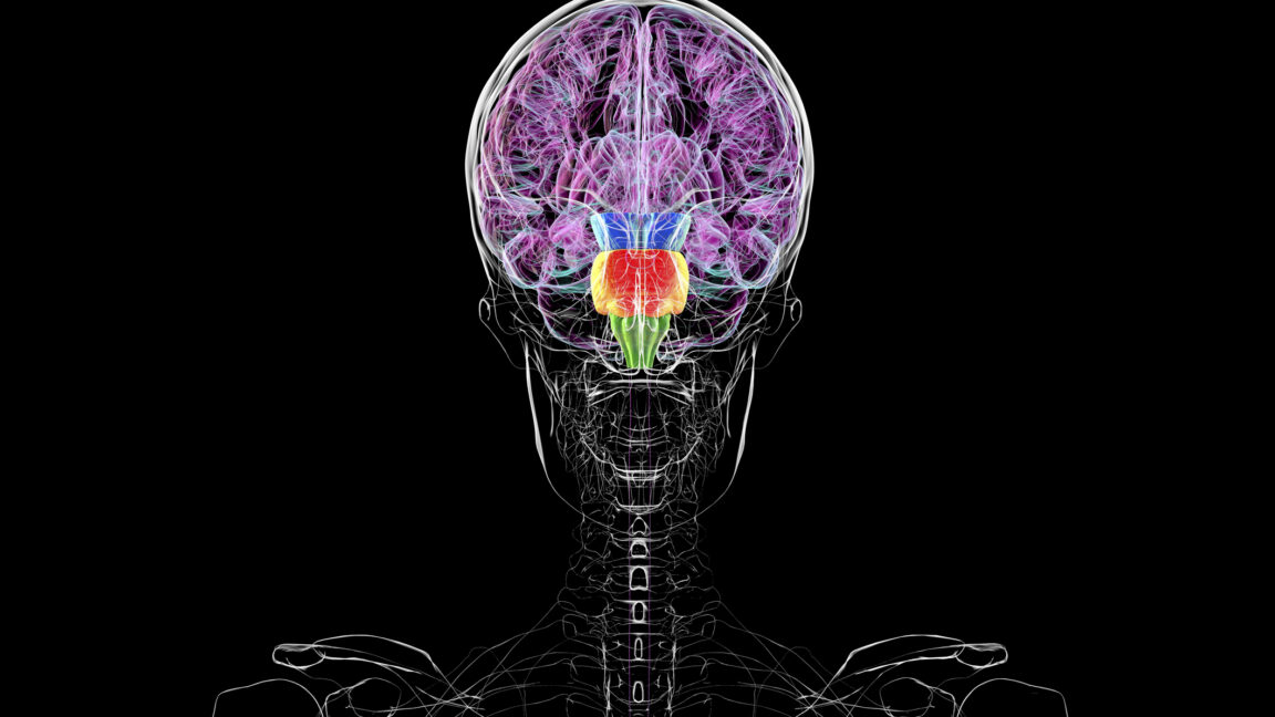
Late last week, we started seeing headlines about how new research was suggesting we might have two brains instead of the one that seems to show up on MRI scans. As is so often the case, these rumors can be traced back to a single press release put out by one of the universities where the work was done.
You do not have two brains. It’s a silly way to look at the results of the new paper.
But there are far better ways to look at the results, ways that can help us understand how the brain gets put together and illustrate some of the general ideas behind developmental biology. So let’s take a look at what the research actually shows and place it in the context of brain development.
Where are you?
You may think body segments are something that show up in things like crustaceans and insects. But our nervous system also develops in segments. Vertebrates have four major ones: the forebrain, midbrain, hindbrain, and spinal cord. (Some of these later develop segments within them, but we’ll skip over those.) The new paper is largely focused on providing part of the answer to an obvious question: how do those segments get there?
That sort of question is the bread and butter of developmental biology, a field that focuses on how organisms start off as a single cell and, through a series of carefully timed and choreographed processes, produce all the tissues found in adults.
To think about the question, we need a bit of context. By the time the first cells that are committed to develop into neurons show up, the vertebrate embryo already knows its head from its tail, and its back from its belly. (We won’t go into how the embryo learns that, but we know a lot about that, too.) The embryo consists of three tissues at the time: the endoderm, which will line our guts, the mesoderm, which will form muscles and bone, and the ectoderm, which will go on to form the skin.
Neural cells form as a thickening of the ectoderm that runs down the center of the embryo from head to tail. The center of these thickly packed cells drops down, while the sides fold up, eventually forming an oval-shaped tube that pinches off from the rest of the ectoderm. All the signals that run up your spine, every sound and shape you process, every thought you will ever have—all of these and more depend on the descendants of these cells.
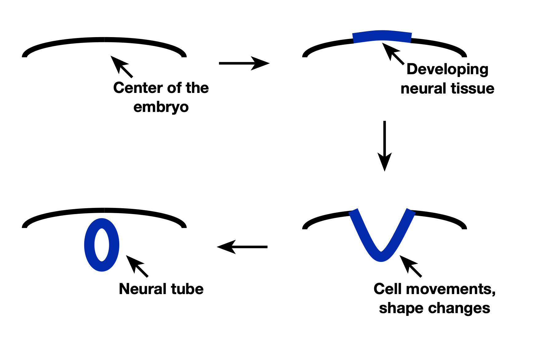
A section across a vertebrate embryo as development proceeds. At first, there’s nothing on top but ectoderm. At the center of the left-right axis of the embryo, the ectoderm thickens and adopts a neural fate (upper right). The neural precursors then start shifting in a way that causes them to adopt a deep V shape (lower right). This process eventually brings the two ends of the developing neural tissue together, and they merge, forming a tube (lower left).
Credit: JOHN TIMMER
A section across a vertebrate embryo as development proceeds. At first, there’s nothing on top but ectoderm. At the center of the left-right axis of the embryo, the ectoderm thickens and adopts a neural fate (upper right). The neural precursors then start shifting in a way that causes them to adopt a deep V shape (lower right). This process eventually brings the two ends of the developing neural tissue together, and they merge, forming a tube (lower left). Credit: JOHN TIMMER
(While this is being presented as a sort of “how vertebrates develop,” there are key differences among them. In mice, the formation of neural tissue happens nearly simultaneously along the entire head-to-tail dimension. In chickens, the process starts in the head and moves slowly to the tail, such that there are already brain structures forming at a time when some of what will be the spinal cord doesn’t even know it will be a nerve cell yet. And tadpoles form a fairly simple spinal cord that gets expanded and reorganized as they change into frogs. So, while the general process is similar in all vertebrates, many species have adapted it to different styles of development.)
By all appearances, all these newly formed neural cells look more or less the same. So we end up back at the original question: how do the segments of the nervous system form?
Remember that, by the time these cells start forming, the embryo already knows its head from its tail. That raises two very simple possibilities. One is that the ectoderm cells that the nerve cells form from already know where they are, and so the nerve cells inherit positional information from them. The alternative is that after the neural cells form, their non-neural neighbors can send signals to them to tell them where they are. So if cells in the head make a different collection of signaling molecules from those in the tail, this can transfer positional information to the developing nervous system, telling it where to form the brain and where to form the spinal cord.
Dividing up the brain
The new work builds on decades of studies that have identified many key regulators of early processes. One of those earlier findings was that the ectoderm of the early embryo activates two genes, one in the front half of the embryo, one in the back. So nerve cells can inherit at least some crude positional information from the ectoderm they form from.
The key to this work is that the researchers modified a copy of these genes so that it activated fluorescent proteins wherever the gene was translated into a protein. So, they engineered mice where half of the early ectoderm glowed red, and the other half glowed cyan. These colors were maintained as the embryo formed nerve cells and the cells started to develop into the brain.
They found that while the hindbrain glowed red, the rest of the brain glowed blue. The inherited positional information set up one of the key boundaries in the brain. In other words, as soon as cells know they’re going to eventually develop into neurons, they know whether they can potentially form part of the hindbrain and not the mid- or forebrain. (This is, roughly, where the idea of “two brains” in the press release comes from.)
Other experiments expanded on this. Similar genetic tools let them activate a fluorescent protein in individual cells in the early ectoderm. They found that, in 96 percent of the cases, the descendants of these single cells were all in the hindbrain, or all in the midbrain and forebrain. They also worked with human stem cells and showed similar things were happening there: Exposure to the right signals would tell the stem cells where they were, and the nerve cells inherited that information as they formed. If you wanted to form neurons that are only found in the hindbrain, you have to first send the stem cells down the hindbrain path.
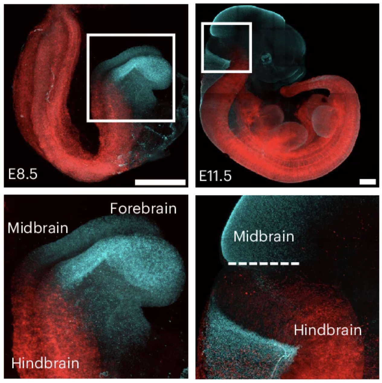
Early cell labeling shows that the hindbrain and spinal cord (red) remain a separate fate from the midbrain and forebrain (cyan).
Credit: Jokhai et. al.
Early cell labeling shows that the hindbrain and spinal cord (red) remain a separate fate from the midbrain and forebrain (cyan). Credit: Jokhai et. al.
There obviously may still be some flexibility here—the 96 percent exclusive behavior they saw is not 100 percent. It’s not clear whether that’s a limitation of their experimental system or just a product of the fact that cells at the border between the front and back of the ectoderm can change fates if they wander a bit in the right direction. Things happen very quickly in early development, and it’s also not clear how the timing of what we can see compares to the timing of what’s going on with the genes that were used to mark different cells.
It’s also less certain what’s going on at the other key borders. Differences between the mid- and forebrain seem to arise later, and largely due to the action of signaling molecules that diffuse through the developing brain and provide positional information. In contrast, very little is known about how the hindbrain and spinal cord adopt different fates; the new paper cites only one reference on the topic. That reference suggests they are also separate fates very early in development, but it seems that more work needs to be done there.
Why does any of this matter?
Obviously, the different areas of our brain and the spinal cord go on to form very different structures, which later perform very different functions. So forming these segments is important. But it’s also important that the different identities get established early—in fact, it’s essential to their ability to develop so differently.
There are two key reasons for this. The first is that having a different identity allows cells to respond to the same signal in different ways. For example, early on when it’s a relatively simple tube, pretty much the entire nervous system is exposed to the same two signals: a molecule called sonic hedgehog diffuses from the bottom of the tube, and a group of molecules called BMPs from the top.
You might expect that, since all the cells of the tube are neural precursors, they’d respond to the same signals in the same ways. But they don’t. Instead, their segment identity directs these signals into overlapping sets of responses that differ in the different areas of the brain. In other words, when midbrain cells see BMPs, they respond in different ways than the cells of the spinal cord do. By dividing the nervous system up early, cells inherit a developmental history that allows a limited number of signals to trigger a great deal of complexity.
(The way that developmental history changes how things are interpreted also enables the signals to be recycled. BMPs initially tell cells whether to develop as neurons or not. Later, they tell developing neurons what identity to adopt once the neuron matures. Later still, BMPs help direct the migration of mature neurons in the developing spine. We don’t have to evolve entirely new signaling systems in order to do different things.)
The other thing is that the junction between segments can be a distinct environment, one where cells are exposed to influences from both of the neighboring identities. At the midbrain-hindbrain boundary, for example, cells activate a distinct set of genes that include signaling molecules that go on to influence the development of the neighboring tissues on both sides of the boundary. As you’d expect from the above, these signals made at the border trigger different responses in the midbrain and hindbrain, since those two segments have different developmental histories.
While the new work says nothing about how many brains you actually have (it’s one with several distinct regions within it), it provides another piece for the puzzle of how the incredible complexity of our brains gets generated from a flat sheet of cells that, just a few hours before, would have happily developed into our skin instead. And it provides a great window into how organismal development works in general and the sorts of experiments we can do to help however many brains we have understand these processes.

John is Ars Technica's science editor. He has a Bachelor of Arts in Biochemistry from Columbia University, and a Ph.D. in Molecular and Cell Biology from the University of California, Berkeley. When physically separated from his keyboard, he tends to seek out a bicycle, or a scenic location for communing with his hiking boots.
KioskNews shows a cleaned-up reading view extracted from the publisher’s page — the original always lives on their site, not ours.