Chinese doctors shrink lung tumour for baby still in mother’s womb, using needle
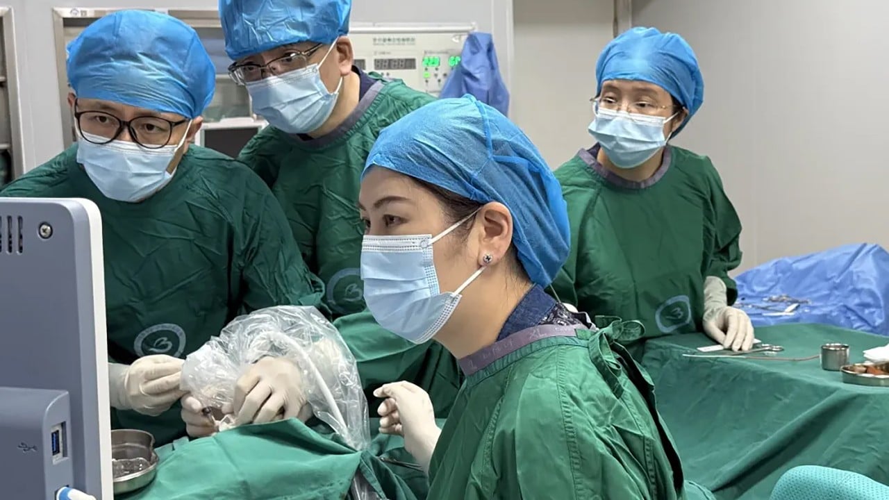
Chinese doctors have performed an unprecedented surgery to save an unborn child who developed a fast-growing tumour.
A medical team from Shenzhen and Hong Kong recently completed a challenging, precise removal of a foetal chest tumour while the baby was still in the womb, offering new hope to families facing similarly dire prognoses.
Jointly performed by doctors from Shenzhen Baoan Women’s and Children’s Hospital and the Chinese University of Hong Kong, the procedure was detailed and published on August 20 in the peer-reviewed journal Ultrasound in Obstetrics & Gynecology.
This marks China “stepping to the global forefront” in the field of minimally invasive interventional treatments for solid foetal tumours, according to the mainland science media platform Advanced Life Science in a September 15 article.
The 40-year-old mother was 22 weeks pregnant when her foetus was diagnosed with microcystic congenital pulmonary airway malformation (CPAM), the medical report said.
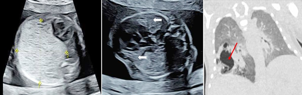
CPAM is a rare congenital condition caused by the abnormal growth of lung tissue during embryonic development. While benign, it is a rapidly growing lung tumour that can lead to heart failure and hydrops, a severe accumulation of fluid.
Scans revealed a solid tumour measuring about 5cm (2 inches) and occupying the foetus’ entire right chest cavity. It severely compressed the heart and lungs and had already caused ascites, a dangerous build-up of fluid in the abdomen.
Standard treatments usually involve draining large fluid pockets or cutting off a tumour’s main blood supply. However, because this specific growth consisted of tiny cysts and lacked a central blood vessel, traditional methods were ineffective.
To save the infant, the medical team adopted a novel approach.
Guided by high-definition, real-time ultrasound imaging, the team precisely mapped a route.
They smoothly inserted a fine ablation needle – a thin, specialised medical probe – through the mother’s abdomen, the uterine wall and the foetal chest wall, reaching directly into the core of the tumour.
The microwave technology works by vibrating water molecules to create intense heat, essentially destroying the tumour tissue from the inside out.
The active heating process was exceptionally fast, taking a mere two minutes in total.
Doctors closely watched the heat spread on their monitors to ensure the baby’s healthy organs remained unharmed.
The results were immediate. Just three days after the procedure, the tumour’s volume had decreased significantly and the fluid build-up was completely gone.
The infant was delivered via caesarean section at 37 weeks, weighing 2.9kg (6.4lbs). At birth, the baby achieved a perfect score of 10 on the Apgar scale – a quick test used to check a newborn’s physical condition – indicating excellent overall health.
At four months of age, the infant remains symptom-free and has required no surgical intervention.
According to the Advanced Life Science article, the success of the surgery was not only a highly challenging clinical rescue but also set “a new milestone” in the international history of maternal-foetal medicine.
KioskNews shows a cleaned-up reading view extracted from the publisher’s page — the original always lives on their site, not ours.