Scientists create mice with part-human brains
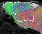
Researchers have created mice with half-human brains in an effort to understand and develop new treatments for disorders such as schizophrenia, epilepsy, cerebral palsy, intellectual disability and rare forms of dementia.
The scientists transplanted lab-grown human brain cells into animals that were engineered to be born without a cortex or hippocampus. This made space for the human tissue to grow inside the rodents’ skulls.
The procedure means scientists can now take cells from patients with brain disorders, turn them into brain tissue in the laboratory, and grow that tissue in living animals. The animals can then be studied to see how the disorder takes hold in human brain tissue, and how drugs might treat the conditions.
“We’ve been trying really hard as a community to find therapeutic solutions for these conditions, but the reality is that in psychiatry and neurology we’ve been left behind [by] every single branch of medicine and we have fewer therapeutics than, again, every single branch of medicine,” said Sergiu Pașca, a professor of psychiatry who led the research at Stanford University.
“That could be because the human brain is very complex, but it’s also because the human brain is inaccessible,” he added. “To a large extent, our goal has been to make aspects of human brain development and function accessible for investigation.”
The work is the latest from the field of neural organoids, where human brain cells, grown in the lab, assemble into tiny but complex structures that mimic some of the features of real brains.
While potentially transformative for brain medicine, organoids have sparked a wave of ethical concerns, not least around whether the clumps of tissue could become conscious or feel pain, and the welfare of animals implanted with organoids.
Pașca said the work had received extensive ethical oversight from the start.
Experts said this would need to continue. Emily Jackson, professor of law at London School of Economics and chair of a recent report on neural organoids for the Nuffield Council on Bioethics, said: “Animal welfare is a really important concern, and it will be necessary to closely monitor these animals in order to evaluate the impact on them.”
In previous work, the Stanford team transplanted human neurons into rat brains. The tissue took root and wired into the animals’ brain circuits, but there was too little room for the human tissue to grow very much.
To solve the problem, the researchers genetically engineered mice to stunt the growth of key brain regions, the cerebral cortex and hippocampus. Surprisingly, the mice survived because the remaining parts of the brain took on new roles. While the mice look normal, they are cautious on their feet and more forgetful.
Writing in Nature, Paşca describes how the mice were injected with human brain organoids that were themselves created by reprogramming donated skin cells. The newborn mice received several injections, each containing about 100,000 human brain cells, into the space where their own brain tissue was missing. In total, the rodents lacked about 14m mouse brain cells and gained about 4m human ones.
Three months after surgery, the human tissue had hooked up to the mouse’s blood supply and almost entirely filled the cavity, taking up about half the size of the rodent’s brain. Some of the human neurons formed connections with mouse brain cells and spinal cord.
The human neurons were not structured or wired up in the same way as in people, and the brain tissue was immature, equivalent to that found halfway through human pregnancy. Tests on the “xenocortical” mice showed the animals were not enhanced by the transplants, but their shaky gait and cognitive problems did improve a little.
To demonstrate how the mice could shed light on human brain disorders, the researchers exposed some of the animals to five hours of low oxygen. This showed how vulnerable human nerve cells are to oxygen deprivation, which in pregnancy and birth can cause cerebral palsy.
The researchers found that human brain tissue in the mice contained rare cells known as von Economo neurons, which have only been seen in postmortem examinations. The cells are among the first to die in frontotemporal dementia, a rare form of the disease, which Paşca now hopes to study in xenocortical mice.
Prof Madeline Lancaster, group leader at the MRC Laboratory of Molecular Biology in Cambridge, said the approach was best suited to questions about disorders and treatments where a whole animal was needed. “It’s less clear to me how this will inform our understanding of human brain development since it is rather artificial, and not at all like how the brain develops naturally,” she said.
Many researchers working on brain organoids are growing them in vitro, or in dishes. The hope is they help to reduce the number of animals used in research. “It’s obviously ethically sensitive and there needs to be a very good reason to do this type of animal experimentation, which most scientific avenues do not require,” Lancaster added. “The field is still aiming for fully in vitro solutions, but this work can also inform on what may be needed to improve those in vitro models to reach more mature stages.”
KioskNews shows a cleaned-up reading view extracted from the publisher’s page — the original always lives on their site, not ours.